[关闭]
您好!欢迎来到炼石商城   请登录  注册    兼职小伙伴  
我的购物车
0
Qiagen 凯杰 Biosharp Omega

首页酶、辅酶及抑制剂MCE>

图片仅供参考,请以实物为准
收藏

Sanguinarine chloride 纯度:96.10% 100mg

价:
0.00
价:
¥0.00

号:HY-N0052A

牌:MCE

账期 货到付款

(预计4-5工作日到货(以MCE官网货期为准))

亲,想了解价格请QQ咨询客服!

工作时间

周一至周五:9:00-18:00

咨询电话

0771-3293894

在线咨询

客服 郭恒 蔡玉坤 曾宪飞 技术咨询

浏览了该商品的用户还浏览了

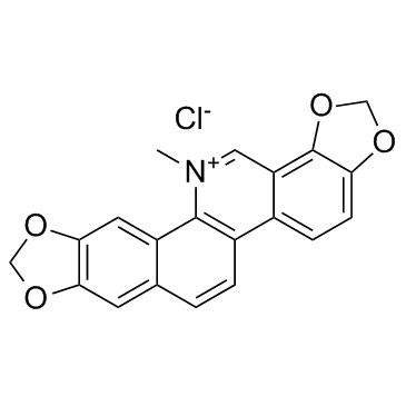
Sanguinarine chloride 是一种来源于 Sanguinaria Canadensis 的生物碱,可通过激活活性氧 (ROS) 的产生来刺激细胞凋亡。Sanguinarine 诱导的细胞凋亡与 JNK 和 NF-κB 的活化有关。

Description

Sanguinarine chloride, a benzophenanthridine alkaloid derived from the root of Sanguinaria Canadensis, can stimulate apoptosis via activating the production of reactive oxygen species (ROS). Sanguinarine-induced apoptosis is associated with the activation of JNK and NF-κB.

IC50 & Target

Apoptosis[1]

In Vitro

Sanguinarine (SANG)-induced apoptosis is associated with the activation of JNK and NF-κB signal pathways.To determine the effects of Sanguinarine on cell viability, 22B-cFluc cells are stimulated with different concentrations of Sanguinarine for 24 h, and then a CKK-8 assay is performed. The treatment with Sanguinarine decreases the proliferation of 22B cells in a dose-dependent manner. Meanwhile, the cytosolic extracts of 22B-cFluc cells treated with different dose of Sanguinarine are measured to detect cellular caspase-3 activity using Ac-DEVD-pNA, which is a validated caspase-3 substrate. The absorbance at 450 nm increases in a dose-dependent manner, indicating increased caspase-3 activity stimulated by Sanguinarine[1].

In Vivo

To evaluate the apoptosis induced by Sanguinarine (SANG) in vivo, 22B-cFluc cells are inoculated subcutaneously into one flank of nude mice and xenograft models are allowed to establish. Mice are treated intravenously with 10 mg/kg of Sanguinarine. At 24, 48 and 72 h after treatment, bioluminescent imaging is performed after i.p. injection of mice with 150 mg/kg of D-luciferin substrate. Sanguinarine treatment induces an obvious increase of luminescent signal as early as 48 h after initial treatment. A sustained bioluminescent imaging (BLI) intensity increased is observed throughout the course of experiment. At 72 h after treatment, the tumors are collected and subjected to TUNEL staining for evaluating apoptosis. Compared with the control tumors, the group treated with Sanguinarine exhibits significantly more cell apoptosis, indicated by the increased green signals from the sporadic apoptotic cells[1].

Solvent & Solubility
In Vitro:  

10 mM in H2O

Preparing
Stock Solutions
Concentration Solvent Mass 1 mg 5 mg 10 mg
1 mM 2.7190 mL 13.5951 mL 27.1902 mL
5 mM 0.5438 mL 2.7190 mL 5.4380 mL
10 mM 0.2719 mL 1.3595 mL 2.7190 mL
* Please refer to the solubility information to select the appropriate solvent.
References
Kinase Assay
[1]

The caspase-3 activity is measured using a caspase-3 activity assay kit. Briefly, the cells treated by different concentrations of Sanguinarine (0.5 μM, 1 μM, 2 μM, 4 μM) or control DMSO are collected, washed and lysed in a lysis buffer for 30 min on ice. The supernatants are then collected by centrifuging at 1,2000 rpm for 10 min. The Ac-DEVD-pNA (2 mM) is added to each sample and incubated at 37°C for 1 h. The optical density (OD) of each sample is finally quantified at a wavelength of 405 nm using a spectrophotometer. The p-NA standard is used to calibrate the caspase-3 activity of each sample[1].

MCE has not independently confirmed the accuracy of these methods. They are for reference only.

Cell Assay
[1]

The cell viability of Sanguinarine is determined by CCK-8 assay using a cell counting kit-8. Briefly, 22B-cFluc cells are seeded in a 96-well plate (5×10³ cells/well) and treated with different concentrations of Sanguinarine (0.5 μM, 1 μM, 2 μM, 4 μM) for 24 h. Then 10 mL CKK-8 is added to each well for 4 h and the absorbance at 450 nm is measured with a microplate reader. The optical density (OD) values are determined to reflect the viable cell populations from each well[1].

MCE has not independently confirmed the accuracy of these methods. They are for reference only.

Animal Administration
[1]

Mice[1]
Xenografted tumor models are prepared by injection of 1×10⁷ 22B-cFluc cells suspended in PBS into nude mouse (n=6). After tumors reach a volume of approximately 100 mm3, Sanguinarine (10 mg/kg) is i.v. injected into mice. After injection for 24 h, 48 h and 72 h, mice are given a single i.p. dose of 150 mg/kg D-luciferin and bioluminescence imaging are performed using a Xenogen Lumina II system. The signal intensity in the region of interest is expressed using the Living Image software 4.1. For the anti-tumor therapy studies, one group of tumor-bearing mice (n=6) receive intravenously 10 mg/kg of Sanguinarine every other day throughout the experimental period, while the control group of mice (n=6) receive DMSO only. Tumor growth measurement is calculated[1].

MCE has not independently confirmed the accuracy of these methods. They are for reference only.

References
Molecular Weight

367.78

Formula

C₂₀H₁₄ClNO₄

CAS No.

5578-73-4

SMILES

C[N+]1=CC2=C3C(OCO3)=CC=C2C(C=CC4=C5)=C1C4=CC6=C5OCO6.[Cl-]

Storage
Powder -20°C 3 years
  4°C 2 years
In solvent -80°C 6 months
  -20°C 1 month
Shipping

Room temperature in continental US; may vary elsewhere

温馨提示:因厂家更改产品包装、产地或者更换随机附件等没有任何提前通知,且每位咨询者购买情况、提问时间等不同,为此以下回复仅对提问者3天内有效,其他网友仅供参考!若由此给您带来不便请多多谅解,谢谢!

我要咨询

咨询内容:
支付方式 货到付款 账期付款

服务热线

0771-3293894